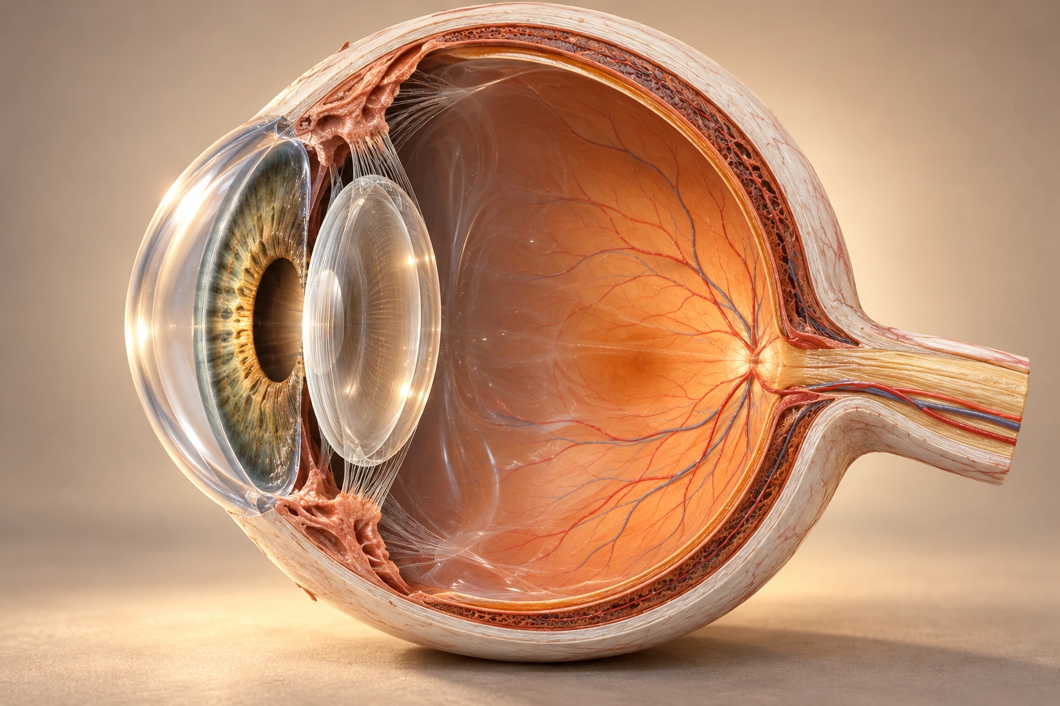
Realistic illustrations of the eye and retina, an interactive model of receptive fields and clickable maps of the visual pathways. No scores or assessments – explore for as long as it helps.
Illustrative anatomical reconstruction, not a photographic observation or a to-scale measurement. Markers identify selected structures; materials and proportions are simplified.Enlarge the retina →
Retina
The retina lines the inside of the eye. There, rods and cones convert light into graded electrical changes. The inner layers of nerve cells then process the information before it leaves the eye.
Text version: the parts of the eye
Cornea
The transparent, curved front surface refracts light as it enters the eye. The cornea accounts for much of the focusing, but does not convert light into nerve signals.
Iris and pupil
The iris is the tissue in front of the lens. The pupil is the opening in the iris, not a black tissue. When the opening changes size, the amount of light admitted changes too.
Lens
The biconvex lens refracts the light further. By changing shape it can focus at different distances – accommodation. Focusing is optics, not transduction.
Vitreous body
A transparent, gel-like mass fills most of the eye's interior. Light passes through the vitreous on its way from the lens to the retina.
Retina
The retina lines the inside of the eye. There, rods and cones convert light into graded electrical changes. The inner layers of nerve cells then process the information before it leaves the eye.
The wall of the eye
Outside the retina lie the vessel-rich choroid and, outermost, the protective sclera. The pigment epithelium lies at the boundary between the neural retina and the choroid.
Optic nerve
The ganglion cells' axons gather and leave the eye in the optic nerve. They carry action potentials towards, among other targets, the LGN in the thalamus. There are no photoreceptors at the exit: that is the blind spot.
Focus or transduction? The cornea and lens refract light so that an image forms on the retina. Only in the photoreceptors is light converted into electrical changes.
Illustrations and models for learning; not measurements.