Chapter 2 · Optional lab
The visual lab
Realistic illustrations of the eye and retina, an interactive model of receptive fields and clickable maps of the visual pathways. No scores or assessments – explore for as long as it helps.
The layers of the retina
Explore the cells
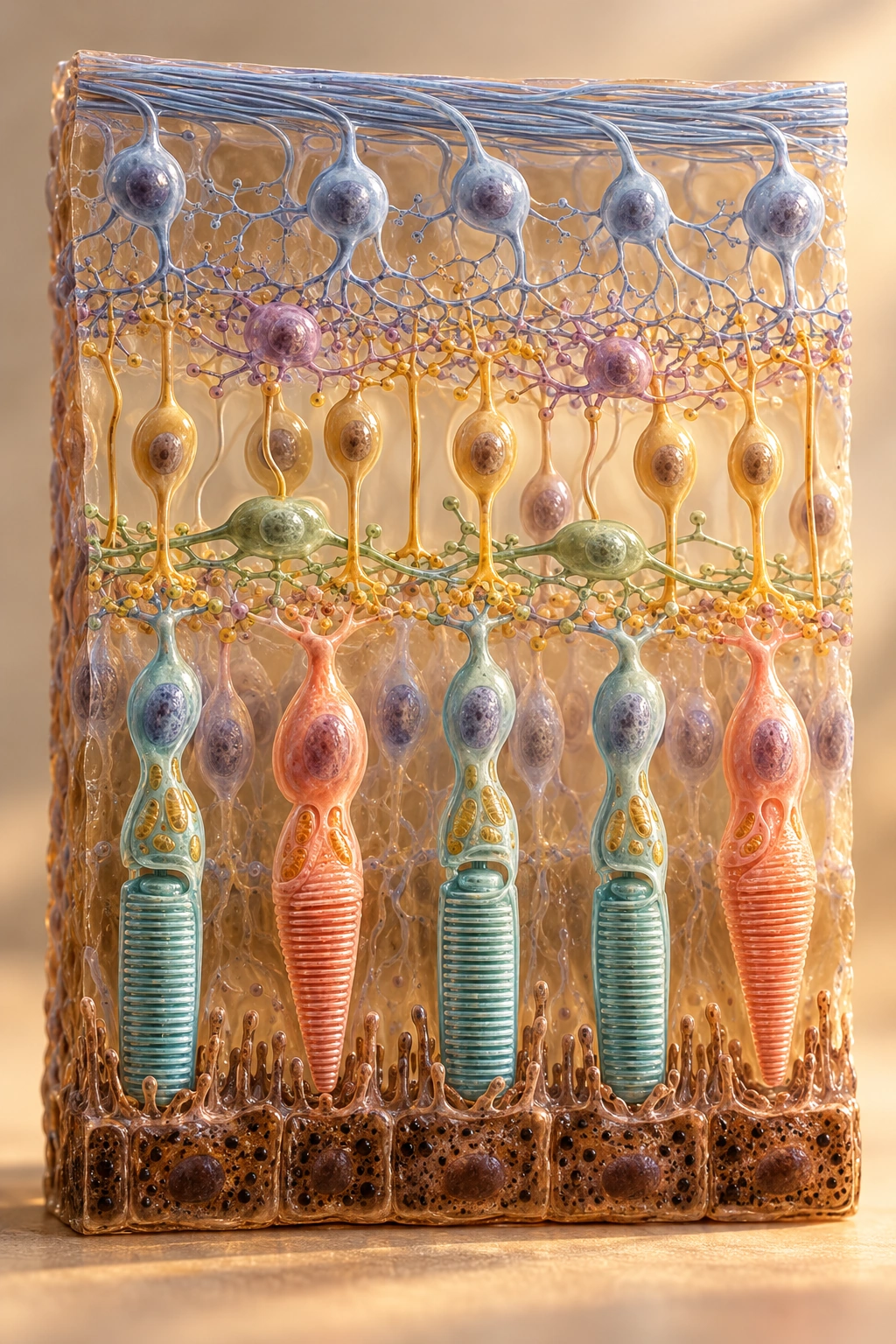
The retina's cells lie in different layers, but their processes meet in the plexiform layers. The visible cells are a selection from a considerably richer network.
Eye interior above · pigment epithelium below
Cones
Cones
Cones have tapering outer segments towards the pigment epithelium and broader synaptic endings in the outer plexiform layer. They provide the basis for colour vision and high acuity in good light. Changed glutamate release affects several types of cone bipolar cells.
Layers from the inside out
- 1
Nerve fiber layer
The ganglion cells' axons run along the inner surface towards the optic nerve.
- 2
Ganglion cell layer
The ganglion cells' cell bodies are shown here.
- 3
Inner plexiform layer
The ganglion cells' dendrites meet bipolar cell terminals and amacrine processes.
- 4
Inner nuclear layer
Bipolar, amacrine and horizontal cell bodies. Amacrine cells lie towards the inside, horizontal cells towards the outside.
- 5
Outer plexiform layer
The photoreceptors' synaptic endings meet bipolar and horizontal processes.
- 6
Outer nuclear layer
The rods' and cones' cell bodies.
- 7
Inner and outer segments
The inner segments lie closer to the cell bodies; the light-sensitive outer segments face the pigment epithelium.
- 8
Retinal pigment epithelium
Supporting cells outside the photoreceptors, next to the choroid.
Two paths, opposite directions
The light must first pass through the retina's inner layers to reach the rods' and cones' outer segments. The main flow of information is then carried back inwards through the network. When light is absorbed, the photoreceptors hyperpolarise and release less glutamate. These are graded changes, not action potentials in the rod or cone. The ganglion cells' axons, by contrast, carry action potentials out of the eye.
The rods' main pathway in dim light includes rod bipolar cells and AII amacrine cells, which relay information to the cone pathways' ON and OFF circuits. That pathway is not drawn here; the rods should therefore not be read as directly connected to the ganglion cells.
In depth: from photopigment to glutamate
When the photopigment absorbs light, a reaction chain begins that reduces the amount of cGMP. cGMP-gated ion channels close, and the photoreceptor hyperpolarises. Lower calcium inflow at the synaptic ending reduces glutamate release. The next cell's response depends on its receptors and on whether it belongs to an ON or an OFF pathway.
Illustrations and models for learning; not measurements.