Skip to content
Cognitive neuroscienceThe Neural Mind
Contents and reading path

Chapter 2

Sensation, Perception and Action

  1. 2.1How does the world become an experience?
  2. 2.2From stimulation to nerve signals
  3. 2.3Vision and visual perception
  4. 2.4Hearing and the direction of sound
  5. 2.5When the senses work together
  6. 2.6The cup in a noisy café
  7. 2.7How does a movement take shape?
  8. 2.8Prediction, feedback and learning
  9. LabThe visual lab

Perception and motor learning – in preparation

Reading path
Coming chapters (planned content)
  1. 1. Introduction to Cognitive Neuroscience
  2. 3. Attention
  3. 4. Memory and Learning
  4. 5. Language
  5. 6. Thinking and Executive Functions
  6. 7. Decision-Making
  7. 8. Emotion and Motivation
  8. 9. Social Cognition
  9. 10. Metacognition and Consciousness

Chapter 2 · Section 4

Hearing and the direction of sound

Hearing sorts sound by frequency in the cochlea – but where the sound comes from must be worked out by comparing the ears.

Sound is pressure variations in the air. When they reach the ear they set the eardrum vibrating, and the small bones of the middle ear – the ossicles – carry the motion on to the cochlea in the inner ear. There the motion propagates through the fluid and sets the basilar membrane in motion. The hair cells on the membrane convert the mechanical influence into changed receptor potentials, which in turn affect the action potentials in the auditory nerve's fibres.
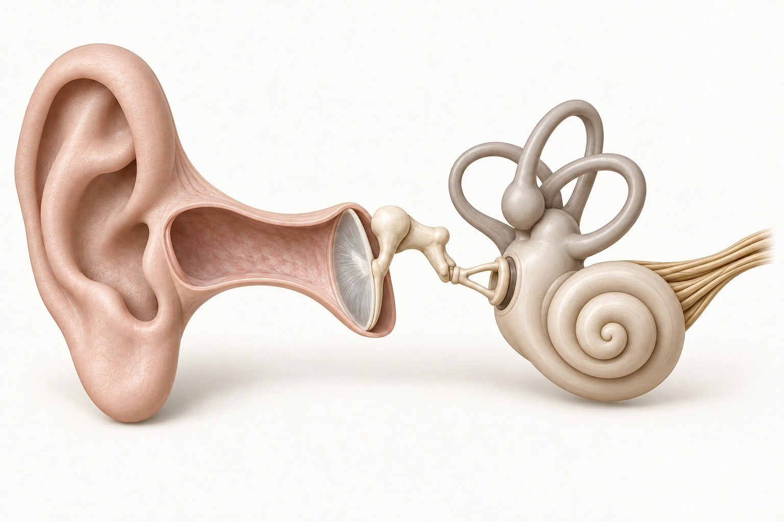
Isolated model of the ear in warm natural colours. On the left are the pinna and ear canal, in the middle the eardrum and the three ossicles, on the right the inner ear with semicircular canals, cochlea and auditory nerve fibres.1Pinna2Ear canal3Eardrum4Ossicles5Cochlea6Auditory nerve7Semicircular canals

Cochlea

The spiral-shaped cochlea is filled with fluid. The motion of the stapes sets the fluid in motion, and along the basilar membrane inside the cochlea the hair cells convert the mechanical influence into changed receptor potentials.

Figure 2.5a. The parts of the ear. Isolated, simplified model. Relative proportions are adjusted for clarity. Choose a marker in the image or in the list below it to read about each part.Pedagogical reconstruction, not anatomical evidence or a to-scale depiction.

The basilar membrane responds differently along its length. Near the base, by the middle ear, it is narrow and stiff and vibrates most for high frequencies; towards the apex it becomes wider and more compliant and responds most to low ones. This ordered frequency map is called tonotopy and is preserved through large parts of the auditory system. But unlike the retina, where the position on the sensory surface corresponds to the position in the visual field, the place in the cochlea says nothing directly about where the sound comes from.
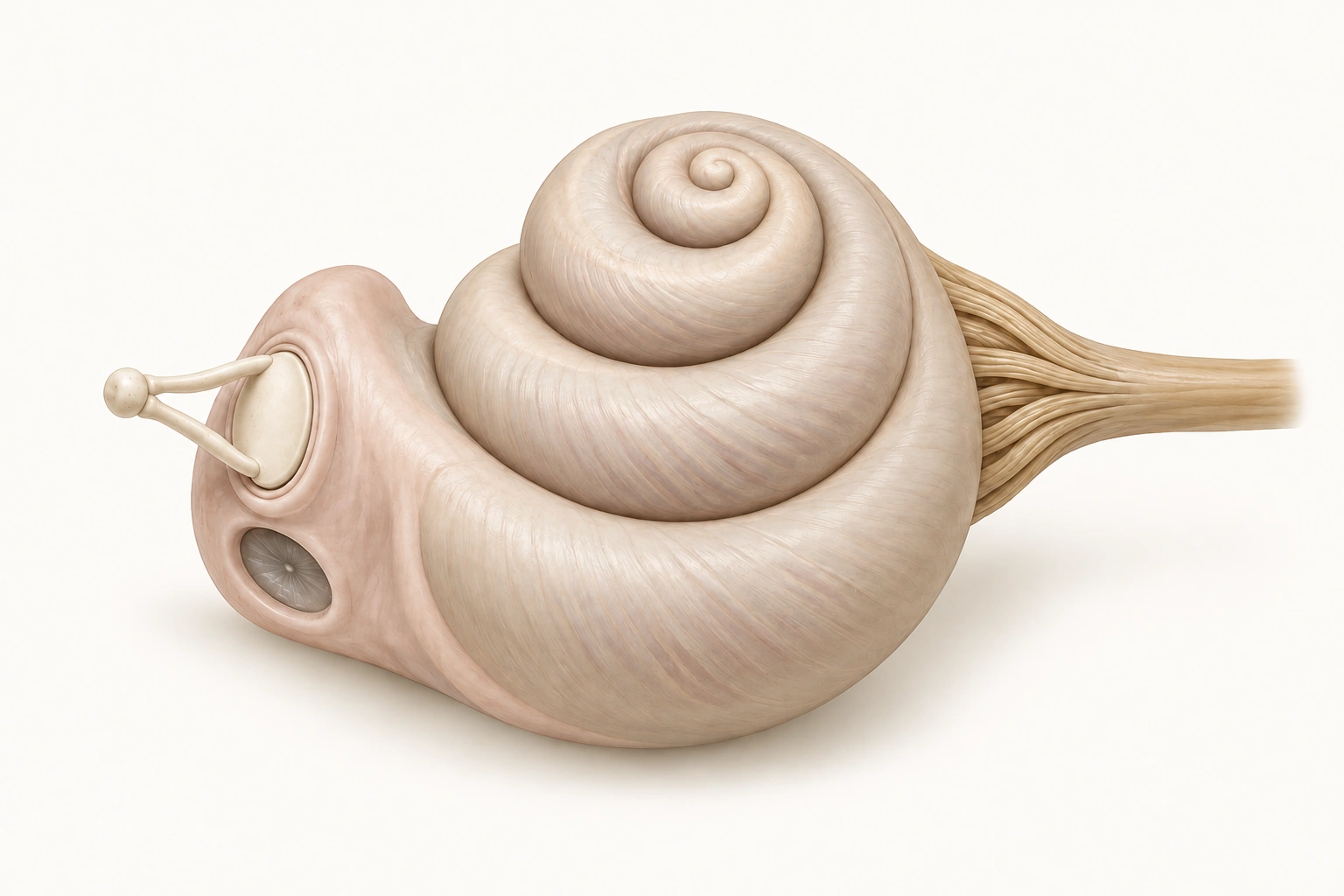
Enlarged isolated model of the cochlea in warm natural colours. On the left is the stapes footplate against the oval window with the round window below, in the middle the spiral cochlea and on the right the auditory nerve fibres.1Stapes and oval window2Base of the cochlea3Apex of the cochlea4Auditory nerve fibres

Base of the cochlea

The base is the first part of the spiral, nearest the middle ear. Inside the cochlea the basilar membrane is narrow and stiff here. High frequencies produce the greatest movement near the base.

Figure 2.5b. The cochlea enlarged. Isolated, simplified model. Relative proportions are adjusted for clarity. Base and apex refer to position along the spiral; tonotopy concerns the basilar membrane inside the cochlea, not its outside.Pedagogical reconstruction, not anatomical evidence or a to-scale depiction.

To see how the frequency ordering lies along the membrane, we unroll the cochlea in a separate functional schematic – a different kind of picture from the model above.

Unrolled cochlea with tonotopyThe basilar membrane drawn as a band from the base on the left, near the middle ear, to the apex on the right. The base is narrow and stiff and responds mainly to high frequencies; towards the apex the band widens and responds to lower frequencies.Basenarrow, stiff – high frequenciesApexwider, flexible – low frequenciesThe basilar membrane unrolled (schematic)

Sound source to the left of a head seen from aboveA head seen from above with left and right ears. A sound source to the front left. The path to the left ear is shorter; the path to the right ear is longer and goes around the head. The sound therefore reaches the left ear slightly earlier and slightly stronger.Sound sourcenose (forwards)Left earRight earshorter pathlonger path around the headTime difference (ITD) and level difference (ILD) between the ears
Figure 2.5c. Tonotopy and two ears. Top: the cochlea unrolled from base to apex. Choose high or low frequency to see where the membrane moves most. Bottom: a sound from the left reaches the left ear first and slightly stronger. No sound is played and this is not a hearing test.Original schematic illustration.

The direction must therefore be computed. Nerve cells in the brainstem compare the signals from the two ears: a sound from the left reaches the left ear slightly earlier (interaural time difference) and can be stronger there, because the head shadows the far ear (interaural level difference). The shape of the outer ear also filters sound differently depending on direction, which helps us tell front from back and up from down.

From the brainstem, several parallel pathways run, with connections on both sides, via the inferior colliculus and the medial geniculate nucleus (MGN) in the thalamus to the auditory cortex in the temporal lobe. It is thus not a single obligatory chain, but a network in which different properties of the sound – frequency, timing, level and direction – are processed in parallel.

You can compare how receptors in vision and hearing convert their stimulation in the receptor comparison in section 2.2, and the descending control that reaches the cochlea through the brainstem is described under feedback in the same section.

Inner and outer hair cells: different contributions

Inner hair cells release glutamate onto the nerve fibres carrying most cochlear afferent signals. Outer hair cells help amplify and shape the mechanical motion. Descending olivocochlear pathways through the brainstem reach different targets: MOC contacts outer hair cells, while LOC influences auditory nerve endings near inner hair cells. There is no direct axon from auditory cortex to a hair cell.

This influence works alongside selection at several brain levels. Animal experiments show peripheral influence from cortex; how much voluntary attention changes human cochlear responses is a more specific question. Compare visual and auditory feedback in section 2.2 →

Sources for this section
  • Scientific referencePurves, D. et al. (2001). Neuroscience (2nd ed.). The Auditory System – Summary.Tonotopy in the cochlea and auditory pathways. Open the source ↗ (new tab)
  • Scientific referencePurves, D. et al. (2001). Neuroscience (2nd ed.). Integrating Information from the Two Ears.Interaural time and level differences. Open the source ↗ (new tab)
  • Scientific referenceGuinan, J. J., Jr. (2010). Cochlear Efferent Innervation and Function. Current Opinion in Otolaryngology & Head and Neck Surgery.Review of olivocochlear systems: MOC to outer hair cells, LOC to auditory nerve fibres near inner hair cells. Open the source ↗ (new tab)
  • Scientific referenceDragicevic, C. D., et al. (2015). The olivocochlear reflex strength and cochlear sensitivity are independently modulated by auditory cortex microstimulation. Journal of the Association for Research in Otolaryngology, 16, 223–240.Cortical microstimulation in chinchillas: peripheral auditory influence through descending systems. MOC contacts outer hair cells; LOC auditory afferents. Not a measure of voluntary human attention. Open the source ↗ (new tab)
  • Figure provenance and model limitations are described on the sources page. About sources and figures